Researcher Spotlight: Shari B. Brosnahan, MD

Dr. Brosnahan helps to lead RECOVER studies that use medical imaging to understand how specific changes in the lungs, heart, and brain can cause Long COVID to affect each person differently.
Like many healthcare providers, RECOVER researcher Shari Brosnahan, MD (NYU Langone Health), was profoundly impacted by the pandemic. “Being in New York, I was part of the first major US surge in COVID-19 cases in early 2020,” she said. “As a doctor working in an intensive care unit (ICU), I felt like it was important to focus my attention on this emerging disease.”

Dr. Brosnahan was well positioned to care for people hospitalized with life-threatening symptoms of COVID-19. She is a specialist in pulmonary medicine (a medical specialty focused on the lungs and respiratory system) and critical care medicine (a medical specialty dedicated to caring for the sickest patients, including those who require a breathing machine). Additionally, she focuses her practice on helping patients who develop blood clots in their lungs.
Whether blood clots form in the lungs or move into the lungs from elsewhere in the body, such as the arms and legs, they can cause chest pain, shortness of breath, fainting, and even death. If one of these clots gets stuck in the blood vessel (artery) that carries blood from the heart to the lungs, it can create a blockage called a pulmonary embolism. If left untreated, a pulmonary embolism can cause the oxygen in the lungs to drop to dangerously low levels, which can lead to permanent lung damage or heart failure.
People with COVID-19 can experience a higher risk of developing a pulmonary embolism, and this elevated risk can last for months after an initial SARS-CoV-2 infection. “It’s clear that COVID-19 can cause large amounts of inflammation (swelling) in the body, which can result in the increased formation of blood clots,” Dr. Brosnahan explained. “RECOVER hopes to help solve why this increased risk can continue after an acute (initial) SARS-CoV-2 infection subsides.”
In addition to pulmonary embolisms, Dr. Brosnahan also studies post-intensive care syndrome (PICS)—a health condition that, like Long COVID, is associated with physical, mental, and emotional symptoms that can last for months following an illness. She brings all of this experience and expertise to several different RECOVER studies.
Through her work on RECOVER, Dr. Brosnahan leads, co-leads, or contributes to multiple studies that use medical imaging to further our understanding of and to increase our ability to diagnose, prevent, and treat Long COVID. Medical imaging refers to various techniques for examining parts of the body that cannot be seen by the naked eye. Medical imaging techniques include X-rays, computed tomography (CT) scans, ultrasounds, and magnetic resonance imaging (MRI). These techniques can reveal details about the structure of and processes taking place within organs, bones, and deep tissues.
According to Dr. Brosnahan, RECOVER’s efforts to learn more about Long COVID from medical imaging began with making sure all researchers collect images in the same way. “I started on RECOVER almost 4 years ago by helping to standardize the testing done across all study sites,” she said. “I helped write the standard operating procedures, or SOP, for all the different imaging tests the sites do, including CT scans and MRIs. This process allows us to learn more about the complexities of Long COVID because it makes it easier for us to compare images taken from different people at different study sites.”
RECOVER is collecting CTs of the chest, MRIs of the brain and heart, exercise tests, echocardiograms (ultrasounds of the heart), and other forms of medical imaging from people taking part in the initiative’s adult and pregnancy observational studies. To date, RECOVER has collected 9,000 images, with the goal to collect over 11,000 images from adults taking part in RECOVER studies.
Each medical image that RECOVER collects undergoes a thorough and detailed research-level scan at study sites called reading centers. With these scans, researchers look for changes in many different small areas of the brain, heart, and lungs.
The rich data these images contain could tell researchers much more about what causes specific symptoms of Long COVID. For example, 2 people with Long COVID may experience the same symptom, but each person’s symptom could have a different cause linked to a unique change that has occurred in their body.
“We’ve spent the first part of RECOVER identifying symptoms and what makes Long COVID a syndrome. What we have to do now is determine what has changed in the body to cause those symptoms and how we can help ease that change,” shared Dr. Brosnahan. “We want to be able to say to a patient that COVID has affected them in this way and that the specific symptom they’re experiencing seems to be more related to changes we observed within their lung architecture, or the way that they exhale during a breathing test, or the size and shape of their brain.”
Currently, the imaging studies focus on analyzing and publishing findings from what Dr. Brosnahan describes as “baseline scans” for CTs of the lungs, MRIs of the brain, and the results of lung function (breathing) tests. This baseline scan is the first medical image taken of a RECOVER study participant. As Dr. Brosnahan explained, this baseline scan records changes in the body that may have already happened or begun happening. Because some studies collect multiple images over time, the baseline scan also sets a reference point that allows researchers to track changes in a study participant’s body as those changes evolve over time.
Dr. Brosnahan emphasized that this standardized, systematic approach to collecting and analyzing medical images is key to finding answers to important questions about Long COVID. “Exposure to the SARS-CoV-2 virus can change your body in many different ways,” she said. “If Long COVID were one disease—if everyone’s organ systems changed the exact same way in response to COVID-19—our research would be very different. What we don’t want to do is to generalize.”
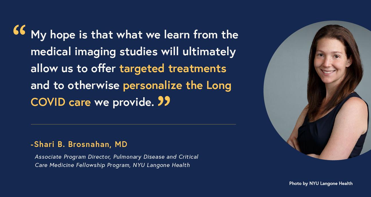
Ultimately, Dr. Brosnahan believes contributing to RECOVER represents an important first step in helping people like the patients she cares for. “My hope,” she said, “is that what we learn from the medical imaging studies will ultimately allow us to offer targeted treatments and to otherwise personalize the Long COVID care we provide.”